|
Introduction The term “nano” comes from the Latin nanus, meaning “dwarf”. This kind of technology is supported in physics, chemistry, materials science, biotechnology, and biosciences, among others, to achieve a multidisciplinary understanding of the phenomena that occur in the materials [1]. Nanotechnology, a relatively new area of study and research, is the “design, characterization, production and application of structures, devices and systems, by controlling shape and size at the nanometer scale”. The particle matter usually ranges from 1 to 100 nm in size. A nanometer (nm) is a billionth of a meter, 10-9. Within this range, materials may have properties considerably different from those expected when they have larger dimensions. Nanoscience depends on the fundamental properties of nano size objects [2]. Nanoparticles are an important class of nanomaterials studied as nanotechnology [1]. Biosynthesis of nanoparticles is becoming a hot topic across the world; this field encompasses a wide spectrum of applications. It plays a critical role with its vast application in nanomedicines, chemical sensing, drug delivery, data storage, cell biology, textiles, food industries, antioxidants, photocatalytic organic dye-degradation, cosmetics, agriculture, and antimicrobial agents [3]. The metallic nanoparticles are considered the most promising as they possess remarkable antibacterial properties due to their large surface area to volume ratio, which can counteract growing microbial resistance against metal ions, antibiotics, and the development of resistant strains. Of these metallic nanoparticles, silver nanoparticles stand out, because of their unique properties, such as chemical stability, good conductivity, catalytic, antibacterial, anti-viral, antifungal, and anti-inflammatory activities. They are usually found as components of composite fibers, cryogenic superconducting materials, cosmetic products, food industry, and in electronic components. Ag nanoparticles have found applications in the biomedical field such as in wound dressings, topical creams, antiseptic sprays, and fabrics. Ag nanoparticles display a broad biocidal effect against pathogenic microorganisms via a disruption of their unicellular membrane, hence, disturbing enzymatic activities. Ag nanoparticles have also been successful in cancer diagnosis and treatment as well [2]. The biological synthesis of nanomaterials has been put into practice since it uses temperature, pH, and pressures that are considered mild conditions. In addition, it has advantages over other synthesis methods, including higher productivity and lower costs. Biological synthesis uses microorganisms and plants (or their extracts). There is a particular interest in using plants because they have many phytochemicals, including ketones, aldehydes, flavonoids, amides, terpenoids, carboxylic acids, phenols, and ascorbic acids. These components are known to reduce metal salts and create metal nanoparticles. However, although there are several studies on nanoparticles, the exact mechanism involved in this process remains uncertain [1]. The plant extracts consist of different bioactive compounds. Phytochemicals sourced from plants assist in the process of conversion of silver ions to AgNPs; the same phytochemicals play dual roles of reducer and stabilizers agents. Concentration of these phytochemicals determines the size, shape, and surface properties of the AgNPs, which defines their functional properties and potential uses [4]. Commonly known as coniferous plants, junipers belong to the genus Juniperus of the family Cupressaceae The genus Juniperus is a monophyletic that consists of almost seventy species [5]. Juniperus is the third largest genus among the conifers found in the world. Plants belonging to this genus grow slowly and can live up to 2000 years. These coniferous plants can be found in all sizes ranging from small flat shrubs to giant forest trees. One thing that makes junipers stand out among other plants is that they can survive in different sites enduring extreme and rapid fluctuations in temperature. They can grow in arid places where other plants cannot survive [5]. J. excelsa subsp. excelsa is a medicinal plant that has been used to treat dysmenorrheal, cough, bronchitis and colds, jaundice and tuberculosis, and to induce menses and expel fetuses [6]. Juniper species are especially used in traditional medicine due to their analgesic, diuretic, antibacterial, anti-microbial, anti-inflammatory, and liver-protective effects [7]. In view of the above, the main purpose of this experimental study was to synthesize Ag-NPs fromJuniperus excelsa M.Bieb leaves and seeds extracts in an inexpensive and simple way, and to characterize these nanoparticles at two different concentrations of Ag+ ion.
Materials and Method The study was conducted in the laboratories of Syrian Private University, Atomic Energy Commission of Syria, the National Commission for Biotechnology in Damascus. Preparation of Plant sample The plant of Juniperus excelsa M.Bieb that used in the study was collected in July 2023 from the al-Khusha in mountains of al-Qalamoun, Ras al-Ma'arra village, Yabroud area, Damascus countryside, Syria. The fresh leaves and seeds were separated from the bark and cleared with tap water. Then, they were washed with distilled water thoroughly and dried for 15 days at room temperature in shadow. The leaves and seeds were stored at -20°C until use. Finally, they were crushed into small pieces immediately before use with an electric grinder, where the particle dimensions were less or equal to 0.2 mm. Preparation of Plant Leavs and Seeds Extracts Extraction was done using the method described by Corciova et al. in 2022 with some modification. 25 g of each dried and powder Juniperus excelsa M.Bieb leaves and seeds were extracted with 100 ml of distilled water, with continuous stirring by a magnetic stirrer at 6 rpm for one hour (at room temperature). Then, it was heated to 60°C for another hour with the same conditions. The alcoholic extract was prepared in the same way, replacing ethanol with distilled water. Each mixture was soaked and left to stand for 24 hours at 4°C and then filtered using Whitman No. 1 filter paper. After that, each supernatant was evaporated using a vacuum rotary evaporator to concentrate the extracts [8]. Preparation Silver Nitrate (AgNO3) Solution The mother solution was prepared from silver nitrate 1M, by dissolving 33.974 g of it in 200 ml of deionized water. Then two dilute solutions were prepared to obtain concentrations of 0.08 and 0.16 M from the mother solution. The containers were kept away from light to prevent oxidation of the silver ions. Synthesis and Characterization of Silver Nanoparticles The synthesis of nanoparticles (Ag-NPs) was carried out by adding 10 mL from the stock solution of each extract to 90 mL for each concentration of AgNO3 aqueous solutions, separately, in a 250 mL flask. The addition was conducted slowly at 25°C for 4 hours with continuous stirring by a magnetic stirrer. While stirring, the color change was monitored where the reaction color transformed from light yellow to dark brown, which indicates the time-dependent formation of Ag-NPs. Afterward, no further color transformation was perceived until the end of the reaction. After that, the pH of the solution adjusted to a value of 7. The solutions were left to stand for 24 h, then were filtered with Whitman No. 1 filter paper. The obtained product was dried at 60°C for 8 h in an oven. Finally, a black powder (nanoparticles) is achieved. They were removed after weighing and transferred to a closed container and kept for further analysis [9]. Characterization of AgNPs The characterization of synthesized nanoparticles was carried out as follows, according to the methods described earlier in Baran (2018). The surface structure was visualized by SEM (TESCAN, Czech Republic) at an accelerating voltage of 30 kV, and elemental analysis measurement was done using EDX (EDAX, USA) at an accelerating voltage of 20 kV [10]. Statistical Analyses The data was subjected to one way-ANOVA IBM SPSS software package for Windows (Version 20, SPSS, Inc., Chicago, IL); the statistical significance was evaluated at P≤ 0.05. The results were presented as mean ± standard deviation based on three replications.
Results and Discussion Researchers discovered nanoparticles made from microorganisms and plant extracts are more cost-effective compared to physical and chemical methods and act as reducing and stabilizing agents. The advantages of synthesis from plant extracts include a sanitary working environment, health and environmental protection, less waste, and the most stable products. Nanoparticle properties are determined by the plant extract sources; individual phytochemical combinations in extracts differ substantially depending on the plant source. As a result, changing the extract composition can change the characteristics of AgNPs [3]. For the synthesis of plant-based nanoparticles, the following are necessary:
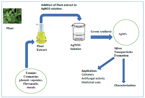
In general, nanoparticle synthesis involves mixing plant extracts with metal precursor salts. They can interact under different reaction conditions (pH, temperature, concentrations, etc.). Here, the formation of nanoparticles takes place in stages. It begins with the set of species equilibrium; once dissolved, the extracts and the precursor salt coordination complexes are formed between the metal ion and the phytochemicals of the extract. The sites where the nanoparticles will grow are created at the second nucleation stage. At the third stage of growth and adsorption, small adjacent nanoparticles come together to form particles of a larger size. Finally, during termination, the final shape of the nanoparticles occurs [1]. The extracts used for nanoparticle synthesis have a wide variety of secondary metabolites involved in nanoparticle reduction, formation, and stabilization. Phytochemicals in the extracts include phenols, flavonoids, alkaloids, terpenoids, proteins, carbohydrates, and amino acids [1]. For biosynthesis, an aqueous AgNO3 solution was mixed with an aqueous and alcoholic Juniperus excelsa M.Bieb extract from leaves and seeds (Figure 1).
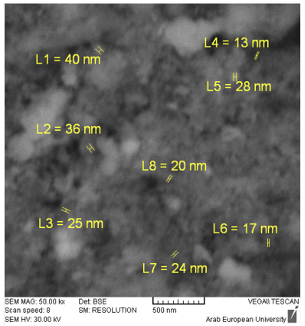
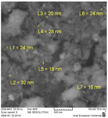
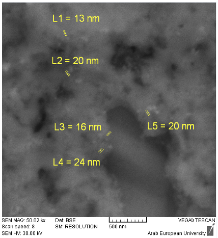
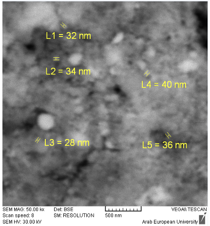
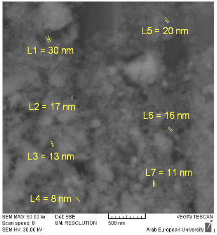
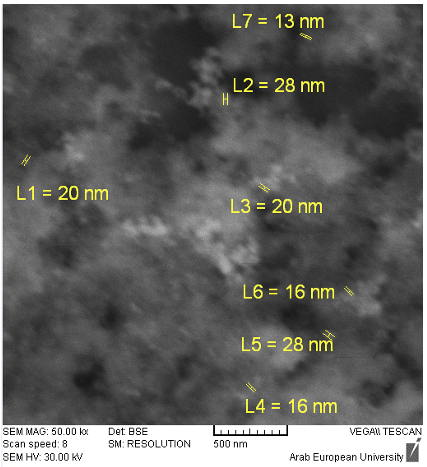
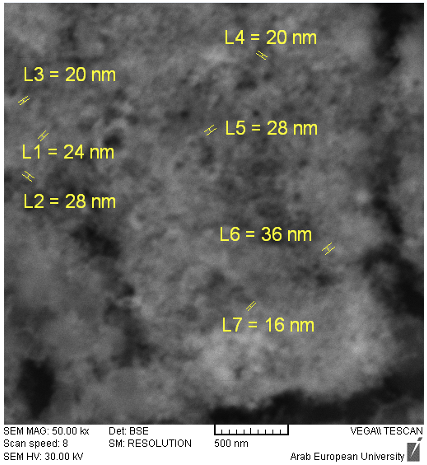
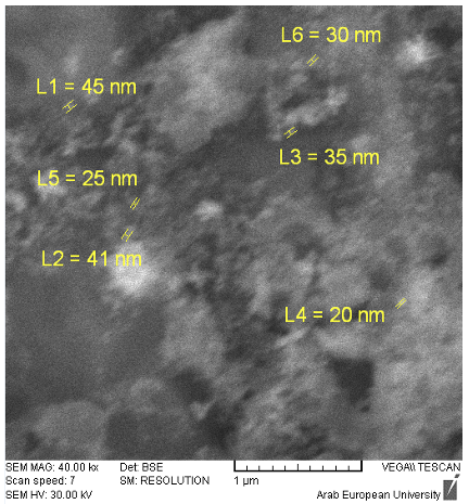
Visual observation showed that the color of the leaves and seeds extracts of Juniperus excelsa M.Bieb, after treatment with Ag precursors, change in color of the reaction mixture from light yellow to dark brown, indicating the formation of AgNPs. This color change is due to quantum confinement and the nanoparticles' dependence on their optical properties [11]. Nanoparticles (NPs) have been the subject of significant analysis due to their ease of preparation, high surface-to-volume ratio, diverse optical properties, unique surface chemistry, and relative simplicity in functionalization compared to their bulk counterparts. The methods frequently used to characterize NPs are UV Visible absorption spectroscopy, SEM (Scanning Electron Microscopy), TEM (Transmission Electron Microscopy), XRD (X-ray Diffraction), DLS (Dynamic Light Scattering), FTIR (Fourier Transmission Infrared Spectroscopy), and EDX (Energy Dispersive X-ray) [12]. In this study, both SEM and EDX were used: Scanning Electron Microscopy (SEM) for providing information about nanoparticle morphology, size, and homogeneity, and Energy Dispersive X-ray spectroscopy (EDX) for estimates of the abundance of elements present over nanoparticles surface. SEM has an ability to contribute morphology and microstructure of bulk as well as nanostructures [13]. Based on the obtained SEM images, the synthesized nanoparticles had various shapes, most of which are irregular and get agglomerated and are multiform. Building blocks of various bioactive reducing agents, lower capping ability of extracts, and H-bonding present in bioactive molecules could be the reason for the agglomeration of the nanoparticles [14]. The average particle size, as calculated using the ImageJ software, from the SEM image was found to be between 13-40 nm, 16-32 nm for aqueous extract of leaves, 13-24 nm, 28-40 nm for alcoholic extract of leaves at 0.08 and 0.16 M, respectively. While was between 8- 30 nm, 13-28 nm for aqueous extract of seeds, 16-36 nm, 20-45 nm for alcoholic extract of seeds at 0.08 and 0.16 M, respectively (Figure 2-9).
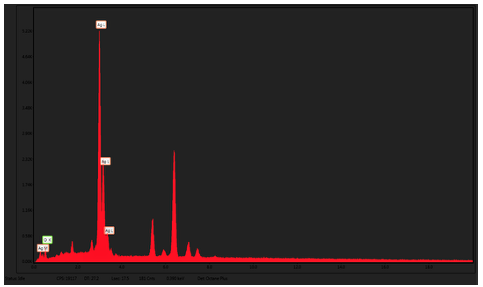
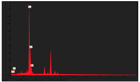
The research studies on the silver nanoparticle synthesis performed showed that the nanoparticles formed ranged in size from 80.11 to 157.01 nm [15], 72.656 nm [16], and 17.3 nm [17]. To use nanoparticles as drug delivery systems, they should be in the range of 10–100 nm. Even though NPs smaller than 10 nm can pass through the nuclear pore and interact with chromosomes and DNA, which is advantageous for gene therapy and diagnostics, they are probably not suitable for drug delivery. For nanomedical applications, the preferred size of nanoparticles is less than 200 nm [18]. In this study, the NPs had a size of less than 100 nm, suggesting the proper size for drug delivery. The previous results show that the lower concentration of silver nitrate (0.08 M) produced smaller nanoparticles than the higher concentration (0.16 M) for all samples studied on leaves and seeds with their aqueous and alcoholic extracts. EDX is an analytical method used to find the chemical compositions of various elements and to determine the relative abundance of particular chemical elements on a solid surface. EDX works on the principle of an interaction of some source of X-ray excitation and a sample. Due to a variance in their atomic structures, every single element proffers the individual assortment of peaks on its electromagnetic emission spectrum. Bombardment of high energy particles on an atom which contains ground state electrons results in the formation of electron-hole by releasing an excited electron from the inner shell. An electron from the outer shell and higher energy shell try to fill the empty shell and this energy disparity between the high-energy shell and the lower energy shell may be dispensed under the form of an X-ray. This could be covered by an energy-dispersive spectrometer. Emitting X-rays helps to identify specific elements and its proportion [10]. Two samples of silver nanoparticles produced from the aqueous extract of the seeds and from the alcoholic extract of juniper leaves were tested for Energy Dispersive X-ray (EDX). In the EDX analysis, it was observed that the content of the element belonged largely to silver. Data analysis revealed that EDX spectra of Ag-NPs mainly contained a specific and intense peak at ⁓3 keV for Ag (88.2% and 88.14%) and for O (11.8% and 11.86%) for the alcoholic extract of the leaves and the aqueous extract of the seeds, respectively, at the concentration 0.08M of silver nitrate. Consequently, the results confirmed the synthesis of Ag-NPs (Figure 10-11).
The studied particles were not observed to have impurities from the hetero-elements of silver and oxygen in the Ag-NPs, so these results indicated that the reaction product was composed of high purity nanoparticles.
Conclusion There are three ways to make nanoparticles: physical, chemical and green synthesis. Green synthesis is the best since it’s very simple, easy to perform, inexpensive, highly efficient, and environmentally friendly. It is the chemical constituents of plants, proteins, carbohydrates, alkaloids, tannins, phenolics, oils and saponins, which can act as reducing and capping agents for NP synthesis. The shape and size distribution of plant-based NP can be controlled by an optimization of reaction conditions, such as temperature, pH, and the amount of plant material. Moreover, the reaction can be scaled up. Silver nanoparticles have proven helpful as nanomedicine, chemical reactions in solar cells, biochemical sensors, and batteries. Ag nanoparticles have found applications in the biomedical field, such as in wound dressings, topical creams, antiseptic sprays, and fabrics. Also, they display a broad biocidal effect against pathogenic microorganisms via a disruption of their unicellular membranes, therefore, disrupting their enzymatic activities. Ag nanoparticles have also been successful in cancer diagnosis and treatment.
Acknowledgment The authors are thankful to the Syrian Private University and Atomic Energy Commission of Syria for providing the facility to conduct the experiments in laboratories.
Funding This research received no external funding.
Conflict of Interest The manuscript was written through contributions of the two authors, and these two authors contributed equally. The authors have given approval to the final version of the manuscript.
References
|
||||||||||||||||||||||||||||||||||||||||